Best Clinical Echocardiogram in Chennai
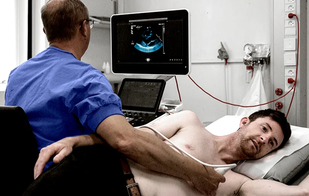
An echocardiogram is a non-invasive ultrasound test that evaluates heart function. During an echo test, high-frequency sound waves from a hand-held wand placed on your chest provide pictures of the heart’s valves and chamber. These echoes are turned into moving pictures of your heart that can be seen on a video screen to evaluate the pumping action of your heart.
Why Is an Echocardiogram Done?
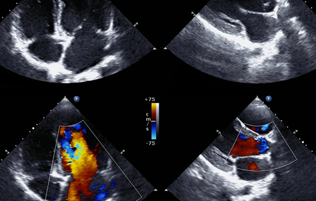
A cardiac echo is used to identify abnormalities in the heart's structure and function. A handheld device sends out sound waves that bounce off your heart and create a moving image of it on a screen. This allows your healthcare provider to look at the anatomy of your heart from many different angles and to observe your heart rhythm.
If you have symptoms of fatigue, shortness of breath, or fainting, you may need a cardiac echo. This is especially true if a stethoscope or an electrocardiogram (EKG) (a test that charts the electrical activity of your heart) suggests that you have a structural heart problem.
Types of Echocardiograms
There are a few different types of echocardiograms. The type that will work best for you depends on a number of factors such as medical conditions you might have and xx
Transthoracic Echocardiogram
This is the standard echocardiogram test. It is similar to the ultrasound tests that are used during pregnancy to view a fetus. This test uses high-frequency sound waves to create a picture of your heart.
Transesophageal Echocardiogram
In people with certain conditions such as a thick chest wall or emphysema, it may be difficult to visualize the heart during an echocardiogram. If you have one of these conditions and need an echo, you might need an invasive ultrasound of your heart known as a transesophageal echocardiogram (TEE). With this, a device is placed in the esophagus in order to view the heart.